Products - Autoimmune - Dermatology -
Bullous autoimmune dermatoses are rare, blister-forming diseases of the outer skin and the adjacent mucous membranes. They are characterized by the formation of autoantibodies against structural proteins of the skin. These structural proteins establish the cell-to-cell contact in keratinocytes within the epidermis and the adhesion of the epidermis to the dermis. Bullous autoimmune dermatoses are divided into four main groups based on their target antigens and the localization of the blisters:
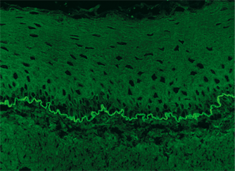 A conclusive diagnosis of blister-forming autoimmune dermatoses requires the detection of both tissue-bound autoantibodies by direct immunofluorescence and circulating autoantibodies. The circulating specific autoantibodies against epidermal antigens (prickle cell desmosomes and epidermal basement membrane) in patient serum are detected using the indirect immunofluorescence test (IIFT) with tissue sections of primary esophagus (or tongue). For further differentiation of autoantibodies against basement membrane structures, tissue sections of primate salt-split skin are used. Final diagnosis is based on a combination of the clinical picture with the detection of autoantibodies against the individual target antigens using IIFT, monospecific ELISA or immunoblot analyses.
Patients who suffer from bullous pemphigoid (BP) exhibit autoantibodies against BP180 and frequently also against BP230. The serum level of autoantibodies against BP180 correlates with the disease activity of BP, the serum level of autoantibodies against BP230 with the duration of the disease. Hence, the Anti-BP180-NC16A-4X ELISA (IgG) and the Anti-BP230-CF ELISA (IgG) are not only suited to reliably serologically identifying BP, but also to monitoring the activity of the disease before and during treatment and assessing the disease duration. Autoantibodies against desmoglein 1 and 3 are markers for pemphigus diseases. IIFT has proven valuable for detecting circulating autoantibodies in pemphigus. ELISA using recombinant desmoglein 1 and 3 offer the same sensitivity and specificity as IIFT. The anti-Dsg1 and -Dsg3 antibody levels measured correlate to a large extend with the severity and activity of the disease and the therapy success. The determination of autoantibodies against envoplakin contributes to diagnosis of PNP as well as differential diagnostic clarification. The determination of autoantibodies against collagen type VII confirms the diagnosis of EBA and enables the delimitation from other bullous autoimmune dermatoses.
For Research Use Only. Not For Use In Diagnostic Procedures.
The individual product regulatory statements may vary, please refer to the instructions for use for more information.
| wdt_ID | Method | Parameter | Substrate | Species/ Antigen |
|---|---|---|---|---|
| 1 | IFA | AB adsorbent (dermatology IFA) |
||
| 2 | ELISA | Dermatology Profile (BP180-NC16A-4X, BP230-CF, desmoglein 1, desmoglein 3, envoplakin, collagen type VII separately) |
antigen-coated microplate wells |
see individual ELISA |
| 3 | ELISA | envoplakin | antigen-coated microplate wells |
recombinant, expression in E. coli |
| 4 | IFA | antibodies against desmoglein 1 (pemphigus-associated) |
||
| 5 | ELISA | desmoglein 1 | antigen-coated microplate wells |
recombinant, expression in mammalian cells, extracellular domain |
| 6 | IFA | desmoglein 1 desmoglein 3 |
transfected cells transfected cells control transfection (3 BIOCHIPs per field) |
EU 90 EU 90 EU 90 |
| 7 | IFA | antibodies against desmoglein 3 (pemphigus-associated) |
||
| 8 | ELISA | desmoglein 3 | antigen-coated microplate wells |
recombinant, expression in mammalian cells, extracellular domain |
| 9 | IFA | antibodies against epidermis: desmosomes (pemphigus vulgaris control) |
||
| 10 | IFA | Dermatology Screen (EM) EUROPattern epidermis: prickle cell desmosomes epidermal basement membrane |
oesophagus | monkey |
| 11 | IFA | epidermis: prickle cell desmosomes epidermal basement membrane |
oesophagus | monkey |
| 12 | IFA | Dermatology Screen 1 EUROPattern epidermis epidermis |
2 BIOCHIPs per field: oesophagus tongue |
monkey monkey |
| 13 | IFA | epidermis epidermis |
oesophagus tongue (2 BIOCHIPs per field) |
monkey monkey |
| 14 | IFA | Dermatology Mosaic 11 epidermis pemphigoid antigens transitional epithelium desmoglein 1 desmoglein 3 BP230gC BP180-NC16A-4X gliadin (GAF-3X) cell nuclei (ANA) endomysium |
11 BIOCHIPs per field: oesophagus salt-split skin bladder mucosa transfected cells transfected cells transfected cells control transfection BP180-NC16A-4X BIOCHIPs gliadin (GAF-3X) BIOCHIPs HEp-2 cells liver |
monkey monkey rat EU 90 EU 90 EU 90 EU 90 human monkey |
| 15 | IFA | Dermatology Mosaic 20 EUROPattern epidermis pemphigoid antigens |
2 BIOCHIPs per field: oesophagus salt-split skin |
monkey monkey |
| 16 | IFA | Dermatology Mosaic 20 epidermis pemphigoid antigens |
2 BIOCHIPs per field: oesophagus salt-split skin |
monkey monkey |
| 17 | IFA | Dermatology Mosaic 7 epidermis pemphigoid antigens BP230gC desmoglein 1 desmoglein 3 BP180-NC16A-4X |
6 BIOCHIPs per field: oesophagus salt-split skin transfected cells transfected cells transfected cells BP180-NC16A-4X BIOCHIPs |
monkey monkey EU 90 EU 90 EU 90 |
| 18 | IFA | antibodies against epidermis: basement membrane (bullous pemphigoid control) |
||
| 19 | IFA | epidermis: prickle cell desmosomes epidermal basement membrane |
tongue | monkey |
| 20 | IFA | antibodies against BP230 (associated with bullous pemphigoid) |
||
| 21 | ELISA | BP230-CF | antigen-coated microplate wells |
recombinant, expression in E. coli |
| 22 | IFA | antibodies against BP180 (associated with bullous pemphigoid) |
||
| 23 | ELISA | BP180-NC16A-4X | antigen-coated microplate wells |
recombinant, expression in E. coli, tetramer of the NC16A domain |
| 24 | IFA | keratin (filaggrin, RA keratin) |
oesophagus | rat |
| 25 | IFA | transitional epithelium (detection of paraneoplastic pemphigus) |
bladder mucosa | rat |
2024 EUROIMMUN US