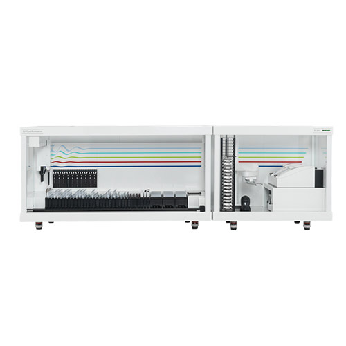
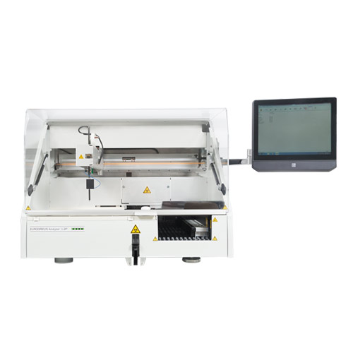
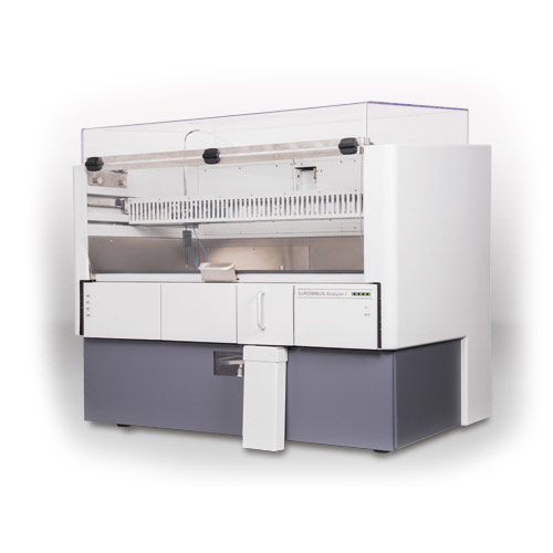
The enzyme-linked immunosorbent assays (ELISAs) from EUROIMMUN use antigens or antibodies coated on a polystyrene plates with 96 wells as a solid phase to bind specific antibodies or antigens in samples through an enzymatic color reaction. The processing can be manual, semi-automated or fully automated. Monospecific ELISAs are used for semi-quantitative or quantitative determination of antibodies or antigens. Semi-quantitative detection of various antibodies on a single microplate strip is achieved using profile ELISAs. Here, the solid phase is coated with an antigen mixture. The antibodies can be detected semi-quantitatively. Afterwards, a differentiated detection must be performed with the respective monospecific assay.
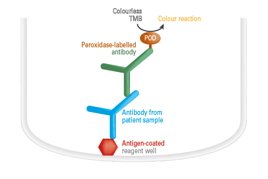 Antibody detection by ELISA
The antigen-coated reagent wells of the microplate are incubated with diluted samples. If the sample contains specific antibodies directed against the antigen, these bind to the antigen-coated reagent wells. In a further step, a peroxidase-labelled antibody (conjugate) is added which binds to the specific antibodies. When the peroxidase substrate tetramethylbenzidine (TMB) has been added, the peroxidase catalyzes a color reaction. The intensity of the resulting color solution is proportional to the antibody concentration in the samples within the measurement range. This can be converted into a concentration by means of a calibration curve in quantitative tests and into a ratio value in semiquantitative tests.
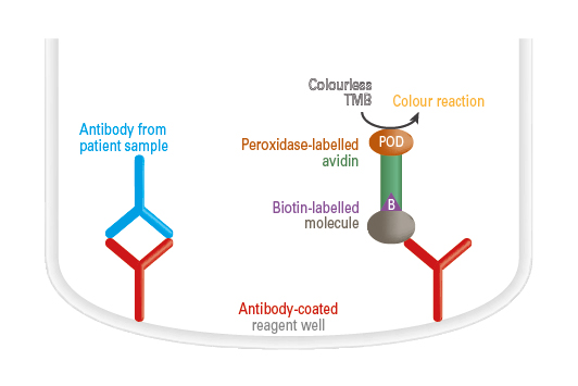 Antibody detection by ELISA (competitive ELISA)
The antigen-coated reagent wells of the microplate are incubated with diluted samples. If a sample contains specific antibodies directed against the antigen, these bind to the antigen-coated reagent wells and inhibit the binding of a biotin-labelled artificial molecule which is added to the reagent well in a subsequent step. Afterwards, peroxidase-labelled avidin is added which binds to the biotin-labelled molecules. In the third incubation step, the peroxidase and the peroxidase substrate tetramethylbenzidine (TMB) catalyze a color reaction. The intensity of the resulting color solution is inversely proportional to the antibody concentration in the sample within the measurement range and can be converted into a concentration by means of a calibration curve in the quantitative tests.
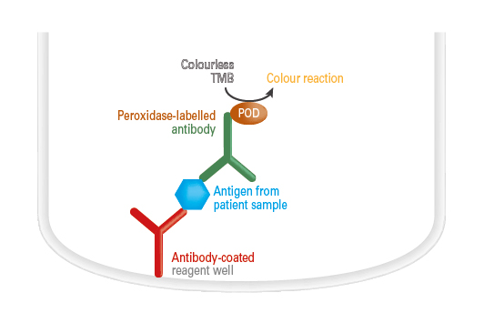 Antigen detection by ELISAs (sandwich ELISA)
The reagent wells of the microplate coated with monospecific antibodies are incubated with diluted samples. If the sample contains the respective antigens, these bind to the antibody-coated reagent well. In a further step, a peroxidase-labelled antibody (conjugate) is added, which binds to another epitope of the antigen. When the peroxidase substrate tetramethylbenzidine (TMB) has been added, the peroxidase catalyzes a color reaction. The intensity of the results color solution is proportional to the antigen concentration in the sample within the measurement range and can be converted into a concentration by means of a calibration curve in the quantitative tests.
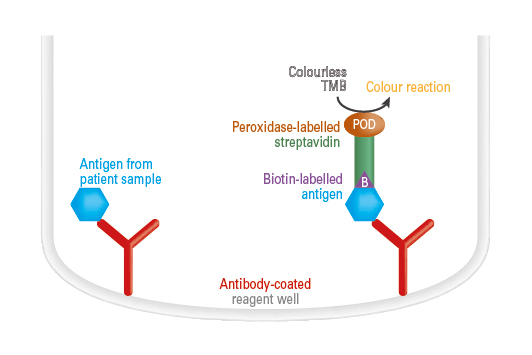 Antigen detection by ELISA (competitive ELISA)
The reagent wells of the microplate coated with monospecific antibodies are incubated with diluted samples and a defined amount of biotin-labelled antigen. If the sample contains the corresponding antigens, these compete with the biotin-labelled antigen for the binding sites in the antibody-coated reagent well. Afterwards, peroxidase-labelled streptavidin is added (conjugate) which binds to the biotin-labelled antigens. When the peroxidase substrate tetramethylbenzidine (TMB) has been added, the peroxidase catalyzes a color reaction. The intensity of the results color solution is inversely proportional to the antigen concentration in the sample within the measurement range and can be converted into a concentration by means of a calibration curve in quantitative tests.
